Canine X-ray Mandible
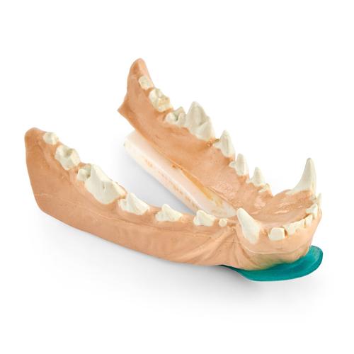
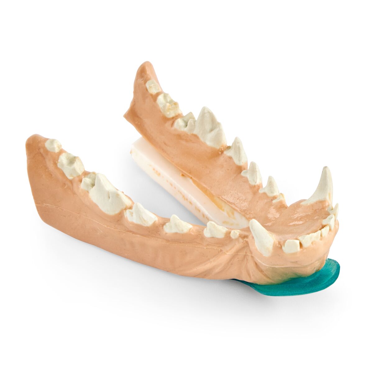
Canine X-ray Mandible
The Canine X-ray Mandible is an essential training model designed to help veterinary students practice radiographic positioning and image capture of the lower jaw. This realistic tool enables learners to produce X-rays that display the detailed anatomy of the mandibular teeth, roots, and surrounding bone structures, supporting a deeper understanding of canine dental health.
A standout feature of this mandible model is its ability to demonstrate the space for the periodontal ligament, which is vital for recognizing dental diseases and conditions. By practicing with this model, students can refine their ability to capture diagnostic-quality radiographs, enhancing both technical precision and clinical confidence.
Uses for the Canine X-ray Mandible:
- Radiographic Positioning: Learn correct positioning techniques for imaging the lower jaw.
- Anatomical Analysis: Examine mandibular teeth, roots, bone structures, and periodontal ligament spaces through clear radiographs.
- Skill Development: Build diagnostic expertise in veterinary dental radiography.
This hands-on model is a valuable addition to veterinary dental training, providing students with practical experience in oral imaging and reinforcing the importance of radiographs in accurate diagnosis.
- 1x Canine X-ray Mandible
Item No.: 1026460
| Weight | 0.1 kg |
| Dimensions | 12 x 5 x 7 cm |
| Brand | VSI |
| MPN: | K9-DENTAL-XRAY-MAN-GR |
- Secure online payment with SSL
- Expert advice
- Easy returns & exchanges
- International shipping available