|
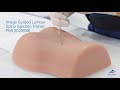
Image Guided Injection Trainer for |
Lumbar Spine |
Thoracic Spine |
Cervical Spine |
|
Product number |
P65 / 1021898 |
P66 / 1021899 |
P67 / 1021900 |
|
Skeletal Anatomy for imaging and palpation |
Sacrum, S1-S3 (with sacral hiatus canal) |
Vertebrae T3-T8 |
Occipital bone (cranial to external occipital protuberance) |
|
Ilium, bilateral without ischial bone and hip joint |
Ribs 3-8 |
Vertebrae C1-T2 |
|
Vertebrae T12-L5 |
|
Ribs 1 and 2 |
|
Coccyx |
|
|
|
Interventional spine procedures |
Transforaminal Epidural Steroid Injections (TFSI) |
Interlaminar Epidural Steroid Injection |
Greater Occipital Nerve (GON) Infiltration |
|
Intralaminar Epidural Iinjection |
Thoracic Transforaminal Injection |
Transforaminal Epidural Steroid Injections (TFSI) |
|
Facet Blocks |
Thoracic Zygapophysial Joint |
Intralaminar Epidural Injection |
|
Medial Branch Block (MBB/RF) |
Nerve (Medial Branch) Injection |
Medial Branch Block |
|
S1-block |
Intraarticular Injection |
Facet radiofrequency denervation |
|
Sacroiliac Joint Injection (SIJ-injection) |
Thoracic Zygapophysial Joint |
C1/C2 intraarticular |
|
|
Intercostal Nerve Block (ICNB) |
Cervical facet intraarticular |
![이미지 유도 경추 주사 트레이너 Image Guided Cervical Spine Injection Trainer P67, 1021900 [P67], 주사실습 및 천자](/imagelibrary/P67/P67_01_이미지-유도-경추-주사-트레이너-Image-Guided-Cervical-Spine-Injection-Trainer-P67.jpg)
![이미지 유도 경추 주사 트레이너 Image Guided Cervical Spine Injection Trainer P67, 1021900 [P67], 주사실습 및 천자](/imagelibrary/P67/P67_02_이미지-유도-경추-주사-트레이너-Image-Guided-Cervical-Spine-Injection-Trainer-P67.jpg)
![이미지 유도 경추 주사 트레이너 Image Guided Cervical Spine Injection Trainer P67, 1021900 [P67], 주사실습 및 천자](/imagelibrary/P67/P67_03_이미지-유도-경추-주사-트레이너-Image-Guided-Cervical-Spine-Injection-Trainer-P67.jpg)
![이미지 유도 경추 주사 트레이너 Image Guided Cervical Spine Injection Trainer P67, 1021900 [P67], 주사실습 및 천자](/imagelibrary/P67/P67_04_이미지-유도-경추-주사-트레이너-Image-Guided-Cervical-Spine-Injection-Trainer-P67.jpg)
![이미지 유도 경추 주사 트레이너 Image Guided Cervical Spine Injection Trainer P67, 1021900 [P67], 주사실습 및 천자](/imagelibrary/P67/P67_05_이미지-유도-경추-주사-트레이너-Image-Guided-Cervical-Spine-Injection-Trainer-P67.jpg)
![이미지 유도 경추 주사 트레이너 Image Guided Cervical Spine Injection Trainer P67, 1021900 [P67], 주사실습 및 천자](/imagelibrary/P67/P67_06_이미지-유도-경추-주사-트레이너-Image-Guided-Cervical-Spine-Injection-Trainer-P67.jpg)
![이미지 유도 경추 주사 트레이너 Image Guided Cervical Spine Injection Trainer P67, 1021900 [P67], 주사실습 및 천자](/imagelibrary/P67/P67_07_이미지-유도-경추-주사-트레이너-Image-Guided-Cervical-Spine-Injection-Trainer-P67.jpg)
![이미지 유도 경추 주사 트레이너 Image Guided Cervical Spine Injection Trainer P67, 1021900 [P67], 주사실습 및 천자](/imagelibrary/P67/P67_08_이미지-유도-경추-주사-트레이너-Image-Guided-Cervical-Spine-Injection-Trainer-P67.jpg)
![이미지 유도 경추 주사 트레이너 Image Guided Cervical Spine Injection Trainer P67, 1021900 [P67], 주사실습 및 천자](/imagelibrary/P67/P67_09_이미지-유도-경추-주사-트레이너-Image-Guided-Cervical-Spine-Injection-Trainer-P67.jpg)
![이미지 유도 경추 주사 트레이너 Image Guided Cervical Spine Injection Trainer P67, 1021900 [P67], 주사실습 및 천자](/imagelibrary/P67/P67_10_이미지-유도-경추-주사-트레이너-Image-Guided-Cervical-Spine-Injection-Trainer-P67.jpg)
![이미지 유도 경추 주사 트레이너 Image Guided Cervical Spine Injection Trainer P67, 1021900 [P67], 주사실습 및 천자](/thumblibrary/P67/P67_01_1200_1200_이미지-유도-경추-주사-트레이너-Image-Guided-Cervical-Spine-Injection-Trainer-P67.jpg)
![이미지 유도 경추 주사 트레이너 Image Guided Cervical Spine Injection Trainer P67, 1021900 [P67], 주사실습 및 천자 (Small)](/thumblibrary/P67/P67_02_1200_1200_이미지-유도-경추-주사-트레이너-Image-Guided-Cervical-Spine-Injection-Trainer-P67.jpg)
![이미지 유도 경추 주사 트레이너 Image Guided Cervical Spine Injection Trainer P67, 1021900 [P67], 주사실습 및 천자 (Small)](/thumblibrary/P67/P67_03_1200_1200_이미지-유도-경추-주사-트레이너-Image-Guided-Cervical-Spine-Injection-Trainer-P67.jpg)
![이미지 유도 경추 주사 트레이너 Image Guided Cervical Spine Injection Trainer P67, 1021900 [P67], 주사실습 및 천자 (Small)](/thumblibrary/P67/P67_04_1200_1200_이미지-유도-경추-주사-트레이너-Image-Guided-Cervical-Spine-Injection-Trainer-P67.jpg)
![이미지 유도 경추 주사 트레이너 Image Guided Cervical Spine Injection Trainer P67, 1021900 [P67], 주사실습 및 천자 (Small)](/thumblibrary/P67/P67_05_1200_1200_이미지-유도-경추-주사-트레이너-Image-Guided-Cervical-Spine-Injection-Trainer-P67.jpg)
![이미지 유도 경추 주사 트레이너 Image Guided Cervical Spine Injection Trainer P67, 1021900 [P67], 주사실습 및 천자 (Small)](/thumblibrary/P67/P67_06_1200_1200_이미지-유도-경추-주사-트레이너-Image-Guided-Cervical-Spine-Injection-Trainer-P67.jpg)
![이미지 유도 경추 주사 트레이너 Image Guided Cervical Spine Injection Trainer P67, 1021900 [P67], 주사실습 및 천자 (Small)](/thumblibrary/P67/P67_07_1200_1200_이미지-유도-경추-주사-트레이너-Image-Guided-Cervical-Spine-Injection-Trainer-P67.jpg)
![이미지 유도 경추 주사 트레이너 Image Guided Cervical Spine Injection Trainer P67, 1021900 [P67], 주사실습 및 천자 (Small)](/thumblibrary/P67/P67_08_1200_1200_이미지-유도-경추-주사-트레이너-Image-Guided-Cervical-Spine-Injection-Trainer-P67.jpg)

![낭종이 있는 유방모형 SONOtrain Breast model with cysts, 1019634 [P124], 초음파 기술 트레이너](/thumblibrary/P124/P124_01_140_140_낭종이-있는-유방모형-SONOtrain-Breast-model-with-cysts.jpg)
![담낭 모형 SONOtrain Gallbladder model, 1019638 [P122], 초음파 기술 트레이너](/thumblibrary/P122/P122_01_140_140_담낭-모형-SONOtrain-Gallbladder-model.jpg)
![낭종이 있는 유방모형 SONOtrain Breast model with tumours, 1019635 [P125], 초음파 기술 트레이너](/thumblibrary/P125/P125_01_140_140_낭종이-있는-유방모형-SONOtrain-Breast-model-with-tumours.jpg)
![이물 모형 SONOtrain Foreign body model, 1019636 [P121], 초음파 기술 트레이너](/thumblibrary/P121/P121_01_140_140_이물-모형-SONOtrain-Foreign-body-model.jpg)
![혈관초음파실습 SONOtrain Ultrasound Vein model, 1019637 [P120], 초음파 기술 트레이너](/thumblibrary/P120/P120_01_140_140_혈관초음파실습-SONOtrain-Ultrasound-Vein-model.jpg)
![이미지 유도 요추 주사 트레이너 Image Guided Lumbar Spinal Injection Trainer P65, 1021898 [P65], 주사실습 및 천자](/thumblibrary/P65/P65_01_140_140_이미지-유도-요추-주사-트레이너-Image-Guided-Lumbar-Spinal-Injection-Trainer-P65.jpg)
![경추 주사 세트, 8000891 [3011957], 시뮬레이션 세트](/thumblibrary/3011957/3011957_01_140_140_경추-주사-세트.jpg)
![요추모형 Lumbar Spinal Column - 3B Smart Anatomy, 1000146 [A74], 척추뼈 모형](/thumblibrary/A74/A74_01_140_140_요추모형-Lumbar-Spinal-Column-3B-Smart-Anatomy.jpg)
![경추 모형 Cervical Spinal Column - 3B Smart Anatomy, 1000144 [A72], 척추뼈 모형](/thumblibrary/A72/A72_01_140_140_경추-모형-Cervical-Spinal-Column-3B-Smart-Anatomy.jpg)
![흉추 모형 Thoracic Spinal Column - 3B Smart Anatomy, 1000145 [A73], 척추뼈 모형](/thumblibrary/A73/A73_01_140_140_흉추-모형-Thoracic-Spinal-Column-3B-Smart-Anatomy.jpg)
![이미지 유도 흉추 주사 트레이너 Image Guided Thoracic Spinal Injection Trainer P66, 1021899 [P66], 주사실습 및 천자](/thumblibrary/P66/P66_01_140_140_이미지-유도-흉추-주사-트레이너-Image-Guided-Thoracic-Spinal-Injection-Trainer-P66.jpg)
![척추 차트, 1001480 [VR1152L], 골격계](/thumblibrary/VR1152L/VR1152L_01_140_140_척추-차트.jpg)
![기본 간호실습 세트 Essential Nursing Lab Set, 8000869 [3011610], 성인간호](/thumblibrary/3011610/3011610_01_140_140_기본-간호실습-세트-Essential-Nursing-Lab-Set.jpg)
![양성 근육 상반신 모형, 31파트 Deluxe Dual Sex Human Muscle Torso Model, 31 part - 3B Smart Anatomy, 1000203 [B40], 인체 상반신 모형](/thumblibrary/B40/B40_01_140_140_양성-근육-상반신-모형-31파트-Deluxe-Dual-Sex-Human-Muscle-Torso-Model-31-part-3B-Smart-Anatomy.jpg)
![피부 모형 (70배 확대) Skin Section, 70 times full-size - 3B Smart Anatomy, 1000289 [J10], 피부 모형](/thumblibrary/J10/J10_01_140_140_피부-모형-70배-확대-Skin-Section-70-times-full-size-3B-Smart-Anatomy.jpg)
![상처치료 및 붕대처치 트레이너 Trainer for wound care and bandaging techniques, 1020592 [P100], 수술봉합 및 붕대감기](/thumblibrary/P100/P100_01_140_140_상처치료-및-붕대처치-트레이너-Trainer-for-wound-care-and-bandaging-techniques.jpg)
![간호 실습 마네킨 베이직 3B Scientific® Patient Care Manikin Basic, 1018817 [P11/1], 성인간호](/thumblibrary/P11-1/P11-1_01_140_140_간호-실습-마네킨-베이직-3B-Scientific-Patient-Care-Manikin-Basic.jpg)
![욕창 치료 트레이너 Decubitus trainer P15, 1019698 [P15], 욕창간호](/thumblibrary/P15/P15_01_140_140_욕창-치료-트레이너-Decubitus-trainer-P15.jpg)
![정맥주사 실습용 팔 P50/1 I.v. Injection Arm P50/1, 1021418 [P50/1], 주사실습 및 천자](/thumblibrary/P50-1/P50-1_01_140_140_정맥주사-실습용-팔-P501-Iv-Injection-Arm-P501.jpg)
![근육주사 시뮬레이터 Intramuscular injection simulator, 1010008 [P54], 주사실습 및 천자](/thumblibrary/P54/P54_01_140_140_근육주사-시뮬레이터-Intramuscular-injection-simulator.jpg)
![카테터 삽관 시뮬레이터 세트 BASIC Catheterization Simulator Set BASIC, 1020842 [P93B-S], 도뇨관 설치](/thumblibrary/P93B-S/P93B-S_01_140_140_카테터-삽관-시뮬레이터-세트-BASIC-Catheterization-Simulator-Set-BASIC.jpg)
![요로(해부생리학)차트, 1001562 [VR1514L], 비뇨기계](/thumblibrary/VR1514L/VR1514L_01_140_140_요로-해부생리학-차트.jpg)
![유방자가검사 모형(착용 가능) Wearable Breast Self Examination Model, 1000343 [L51], 유방 모형](/thumblibrary/L51/L51_01_140_140_유방자가검사-모형-착용-가능-Wearable-Breast-Self-Examination-Model.jpg)
![여성 유방 모형 Model of female breast - 3B Smart Anatomy, 1008497 [L56], 유방 모형](/thumblibrary/L56/L56_01_140_140_여성-유방-모형-Model-of-female-breast-3B-Smart-Anatomy.jpg)